A quiet winter evening (spring, summer, or autumn – whichever you prefer). You come home from work, have dinner, pour yourself a glass of your favorite beer, and settle comfortably on the couch, focused on watching TV.
Suddenly, you feel discomfort in the lower back area, which gradually turns into a dull ache and then into sharp pain. You try to find a body position in which the pain is less intense – no effect. The pain does not subside, you start tossing on the floor or in bed. You are shaking, weakness sets in, nausea, vomiting – no relief. The abdomen is bloated like a drum, gas does not pass. Repeated painful urges to urinate appear.
What is this?! Stale beer?! Fermented cabbage?! No, friends, in urological practice this is one of the most recognizable scenarios – renal colic! What is it? How does it happen? Why? What should be done? How dangerous is it, and what should be done in the first minutes? Let’s sort it out.
What You Need to Know About Renal Colic
What Is Renal Colic?
Renal colic is a collective term that includes several symptoms indicating impaired urine outflow. The pain that occurs belongs to the category of the most intense and ferocious. It is almost impossible to endure, and the patient may even go into shock. It is important to understand that the onset of a pain attack is only a symptom – and a symptom of a condition that is dangerous to the patient’s health and life. As a rule, the occurrence of renal colic indicates urolithiasis and at that moment is associated with the migration of stones from the renal calyx or pelvis into the ureter, but a clinically similar attack is also possible with other causes of obstruction.
Mechanism of Renal Colic Development
As a result of impaired urine outflow, intrarenal pressure increases (intrarenal pelvic pressure, to be more precise) – figuratively speaking, the kidney is “inflated” from the inside. Urine pressure on the parenchyma leads to microcirculatory disturbance and the development of edema. This irritates receptors in the area of the renal hilum and fibrous capsule, which tolerate stretching poorly – this is how the characteristic pain attack forms.
At the consultation, this is clearly reflected in the complaints: the pain “does not depend on body position,” the person cannot find relief, and the wave-like nature of the attack often coincides with attempts of the ureter to “push through” the obstruction.
Who Is Most Often at Risk of Renal Colic
The incidence of renal colic in the population ranges from 1 to 12%. Moreover, more than half of cases occur in men. It should be noted that 13% of cases among all patients with renal colic are caused by other diseases of the kidneys and ureter (tumors, tuberculosis, hydronephrosis, ureterovascular conflict, retroperitoneal fibrosis) and are associated with the passage of blood clots, pus, or mucus that block the lumen of the urinary tract.
That is why the physician’s task is not simply to “relieve the attack,” but to quickly understand what exactly has blocked urine outflow and how safe the situation is.
Causes of Renal Colic and Triggering Factors
Renal colic often occurs after intense physical activity, driving on uneven roads, or excessive intake of fluids or alcohol. Any sudden jolt or overstretching of the urinary tract can “push” a stone and provoke colic.
Behind every such attack there is a cause – most often a mechanical obstruction, inflammation, or spasm that interferes with normal urine outflow. The body reacts instantly: pressure inside the kidney rises, its walls stretch, and the pain becomes almost unbearable. Below are the main causes capable of triggering renal colic.
Urolithiasis
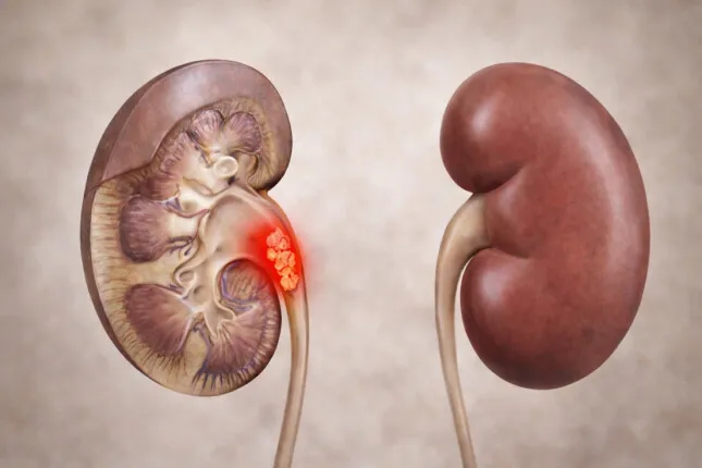
The main cause of renal colic is mechanical obstruction of the urinary tract, most often due to stones. When a stone blocks urine flow, pressure inside the kidney rises sharply, leading to edema, irritation of nerve endings, and sharp “dagger-like” pain. This condition can occur suddenly and catch a person off guard, resembling a storm inside the body.
Inflammatory processes and mucosal edema
In acute inflammatory conditions (pyelonephritis, papillitis), the walls of the ureter become more sensitive to pressure, the ureteral mucosa swells, narrowing its lumen and impairing urine outflow. Accumulation of urine in the renal pelvis creates pressure on the kidney walls and causes a typical pain syndrome. Sometimes colic occurs suddenly – as a reaction to inflammatory edema, even without the presence of a stone.
Anatomical and Congenital Structural Abnormalities
Narrowing of the ureter, congenital bends, or vascular loops compressing it from the outside may cause recurrent pain. Such anomalies often first manifest precisely as renal colic, when urine outflow is impaired during physical exertion or changes in body position.
Tumors and External Compression of the Ureter
Colic may be caused by tumors of the kidney, ureter, or adjacent organs (ovaries, uterus, prostate). A growing mass compresses the ureter, creating an obstacle to urine outflow. In such cases, the pain may be less sharp but progressively increasing, accompanied by a feeling of heaviness and pressure in the lower back.
Traumatic and Postoperative Changes
After surgeries on the pelvic organs, catheter placement, or previous inflammatory processes, scars (strictures) of the ureter may form. Such areas lose elasticity and are easily blocked even by minor edema or sand, triggering an attack of colic. In some cases, pain develops months after the intervention.
Spasm and Functional Disorders of the Ureters
The cause is not always mechanical. Sometimes renal colic occurs due to spasm of the ureteral walls. This may happen with hypothermia, stress, sudden physical strain, or reflexively in response to inflammation of neighboring organs (cystitis, prostatitis). Such spasm also causes acute impairment of urine outflow and a pain attack similar in sensation to that caused by a stone.
Systemic and Metabolic Disorders
Gout, dehydration, excess calcium, vitamin D, or protein-rich food contribute to the formation of salt crystals that deposit in the kidneys and over time form stones. Metabolic disorders often become the background against which the first episode of renal colic occurs – as a signal of developing urolithiasis.
Symptoms of Renal Colic
When an attack begins suddenly, it can be difficult to orient oneself and understand what is happening. In such cases, the main reference points are seven characteristic symptoms of renal colic – their severity and combination help confirm the diagnosis.
Persistent or intermittent pain in the kidney area, “dagger-like pain” in the kidneys
Colic associated with renal pathology is one of the most severe types of pain. The pain may radiate to the groin area and the genital organs (scrotum, penis, vagina, labia) and intensify during urination. Pain may last for several hours or even days, periodically subsiding. The pain is so intense that patients pace around the room, constantly changing position and posture, which usually does not bring relief. This characteristic behavior often allows the diagnosis to be made “at a distance.”
Partial or complete difficulty with urination and passage of gas
When the urinary tract is obstructed by a stone, normal patency and organ tone are disrupted, leading to difficulty urinating. With pronounced spasm, impaired bowel function may also be felt, including difficulty passing gas and bloating.
False urges to defecate and urinate
Irritation of nearby nerve endings and spasm of the pelvic muscles cause false and frequent urges: a person feels an urgent need to go to the toilet, although there may be no real necessity. This is a typical symptom when pathology is localized in the lower urinary tract.
Deterioration of general condition, chills, weakness
Severe pain and possible accompanying inflammation often cause general malaise: chills, weakness, marked lethargy, and decreased work capacity. These symptoms indicate a systemic response of the body and require attention.
Temperature 37–37.5 ℃
Subfebrile temperature (37–37.5 ℃) often accompanies inflammatory processes or a reaction to obstruction. When temperature rises above subfebrile values, the risk of infection and complications increases, requiring urgent medical evaluation. Blood pressure may also rise slightly.
Nausea and vomiting
Severe pain may provoke autonomic reactions – nausea and vomiting, which further worsen the patient’s condition and may require correction of dehydration and non-surgical pain control.
Urine with blood, urine with sediment, and an unusual unpleasant odor
The appearance of pink or red urine indicates injury to the ureteral wall or the renal collecting system by a stone (hematuria). Sediment and an unpleasant odor more often point to a superimposed infection or a large amount of crystals and require immediate laboratory urine testing.
So, How to Help with Renal Colic
Renal colic is one of those conditions where you need to act quickly, but calmly. Panic, abrupt movements, and uncontrolled use of medications only make things worse. The goal of first aid is to relieve pain and avoid harm until the doctor arrives.
Clearly call a doctor immediately (ambulance)
During an attack of renal colic, you must call emergency services right away. But it often takes quite some time for the team to arrive. This is not the clinicians’ fault: traffic jams, weather conditions, an overloaded schedule, and many other factors can prevent a rapid response. That is why the patient and those close to them need to be able to recognize danger and know what can be taken at the peak of pain and what can cause harm.
Provide rest and a comfortable position
Pain during renal colic is so severe that a person instinctively rushes around in search of a position that brings relief. It is best to lay the patient on their back or on the healthy side and try to ensure complete rest and stillness. Sometimes a position with the knees drawn up toward the abdomen helps. Any movements should be smooth and without effort so as not to increase spasm and not to push the stone further along.
Warmth (if there is no fever and no suspicion of inflammation)
If the patient is a stone passer, has long been living with urolithiasis, and this is not their first episode of renal colic, then in the absence of fever you can try placing them in hot water – necessarily in a sitting position. Note that the water should be very hot, as hot as the person can tolerate. A hot bath is contraindicated for patients (especially older ones) with serious cardiovascular disease and those who have had a stroke or heart attack. For such people, first aid for renal colic should involve a hot water bottle placed on the lower back or mustard plasters applied over the kidney area.
Warmth may reduce spasm and subjectively ease pain, but it is not a universal recommendation. Using heat is acceptable only if there is no fever, marked weakness, chills, or other signs of inflammation, and also if there is no suspicion of an “acute abdomen.” If in doubt, do not experiment: in some patients, conditions are hidden under the mask of colic where warming makes the course worse. Otherwise, heating can accelerate an infectious process and worsen the condition.
Pain relief with medications: what is allowed and what is not
If the pain is tolerable until the medical team arrives, it is better to avoid painkillers, because this makes preliminary diagnosis easier. The patient may take 2–3 tablets of No-spa (Drotaverine), one tablet of Ketanov, or another antispasmodic (Papaverine – 1 tablet). If possible, it is better to administer medications intramuscularly (Ketorol, Baralgin) – effectiveness increases several-fold and the effect comes faster. If the listed medications are not available, nitroglycerin can be used (half a tablet under the tongue). It also relaxes smooth muscle and can relieve ureteral spasm.
Nonsteroidal anti-inflammatory drugs, particularly Diclofenac, can effectively relieve pain. I consider intramuscular administration of 75 mg or rectal administration of a suppository at a dose of 100 mg to be optimal. Before using them, it is important to remember contraindications (peptic ulcer disease, bleeding risk, significant kidney problems, allergic reactions), and if in doubt it is better to wait for a doctor. Before the team arrives, it is useful to write down what exactly was taken and in what dose – this speeds up assessment and reduces the risk of repeated administration. If possible, monitor urine for passage of sand or small fragments (it is convenient to collect a portion of urine in a clean container). And even if the attack subsides, do not cancel the call: the pain can return, and temporary relief does not mean urine outflow has fully recovered.
Using painkillers for renal colic is not always necessary or beneficial. Manifestations of urolithiasis may resemble symptoms of other diseases of the abdominal organs and retroperitoneal space. At the same time, the presence of urolithiasis does not exclude the possibility of another acute pathology developing (for example, appendicitis). Therefore, if the attack proceeds atypically, it is better not to do anything until the doctor arrives.
Do Not Try to “Force” the Stone Through
Attempts to “push” the stone are a common and dangerous mistake. Sudden movements, jumping, excessive water intake, or folk remedies can lead to stone impaction, tearing of the ureteral mucosa, and increased pain. If the stone passes on its own, that is luck, but forcibly stimulating this process is категорically unacceptable. The main task is to wait for the doctor and not worsen the obstruction.
Monitoring the Patient’s Condition Until the Doctor Arrives
Heat and antispasmodics can worsen infectious and inflammatory processes in cases of acute appendicitis or other conditions from the group of “acute abdomen” pathologies. That is why it is better to wait for the emergency physician, whose primary task will be to rule out such manifestations of acute abdomen as acute appendicitis, ectopic pregnancy, gallstone disease, peptic ulcer disease, and others, which sometimes must be done together with physicians of other specialties.
To help the doctor determine the cause of the attack more quickly, it is important to remember and, if possible, write down key details: when the pain started, where it is felt, where it radiates, whether there was fever, nausea, or blood in the urine. If urine was collected after the attack, the first portion should be preserved: it may contain sand or small stones, by which the doctor can determine the type of calculi. The more accurate information is provided, the faster a diagnosis can be made and the correct treatment chosen.
Special Cases: Pregnancy and Complications
Special attention should be paid to a patient with renal colic if she is pregnant. Treatment of women with renal colic, especially while pregnant, should be carried out only in a hospital setting. Renal colic in pregnant women is very similar in presentation to labor contractions, so it is important not to delay and to call specialists immediately. Be sure to state right away that the patient is pregnant and indicate the gestational age – the team will arrive faster due to the potential threat to the life of the mother and the baby.
If you have an ovarian cyst, this must be reported to the emergency team immediately. This is important because some gynecological conditions (rupture or torsion of an ovarian cyst) can present with a clinical picture similar to renal colic and require a different approach to emergency care.
When You Should Insist on Hospitalization
If the medications administered by the emergency physician do not bring relief, actively insist on hospitalization in the on-call urology department. It is not a fact that you have renal colic – everyone can make mistakes, especially since the emergency kit does not include a portable ultrasound machine or a biochemical laboratory. As a rule, urology wards are structural units of large medical institutions that also have surgical and gynecological departments. In such a facility, you can be examined more thoroughly with the involvement of related specialists and receive specialized care.
What to Do After an Episode of Renal Colic
If the pain has been relieved, do not relax – the problem is not solved yet! Wait until morning. Wake up (if you managed to fall asleep), wash up, shave – and head straight to the urologist!
Seeing a Urologist After the Episode
How does an outpatient urologist usually proceed? Using additional diagnostic methods, the doctor determines the size of the stone, its location, the degree of upper urinary tract obstruction, and evaluates changes in urine and blood test results. Based on these findings, medical expulsive therapy or other treatment methods appropriate to the clinical situation are prescribed.
Why It Is Important Not to Delay the Visit
It is important to understand that renal colic is only a symptom and most often a sign of a “blockage” in the urinary tract. Obstruction of the upper urinary tract is an extremely dangerous condition in which it is important to eliminate the cause as quickly as possible through appropriate treatment. Otherwise, the patient’s condition may progressively worsen due to secondary infection, increasing intoxication, not to mention the persistent and severe pain syndrome.
In some cases, treatment begins precisely with decompression of the upper urinary tract. Why am I emphasizing timing? During the first hours after the onset of renal colic, an infectious inflammatory process in the kidney often has not yet had time to develop – in other words, the situation is still “uncomplicated.”
When Medical Expulsive Therapy May Be Appropriate
In my subjective opinion, not every episode of renal colic requires immediate active stone removal. In some cases, the doctor may choose a medical expulsive approach – meaning observation, pain control, antispasmodic support, and waiting for the stone to pass spontaneously.
This approach is possible only after examination and diagnostic assessment: when the patient’s condition is stable, there are no signs of significant infection, high fever, worsening urinary obstruction, or other signs of a complicated course. The size and location of the stone also matter, as well as whether it is safe to allow time for spontaneous passage.
It is important to understand: medical expulsive therapy does not mean “just wait.” It is observation within clearly defined time limits and under the supervision of a urologist. If the pain recurs, obstruction worsens, test results deteriorate, or the stone does not pass within the expected timeframe, the treatment strategy is revised in favor of more active intervention.
What to Pay Attention to When Seeking Urological Care
After an episode of renal colic, it is important not only to relieve the pain, but also to see a specialist who understands how to assess such conditions properly, with a clear strategy for urinary tract obstruction and urolithiasis. Below are several practical points that help determine whether the doctor and clinic are prepared for this situation.
- Whether the doctor uses modern diagnostic methods for urolithiasis (ultrasound, X-ray)
- Clinical experience in minimally invasive and endoscopic treatment of urolithiasis
- Clinic equipment – availability of equipment that meets modern European standards for treating patients with urolithiasis (shock wave lithotripter, contact lithotripter, laparoscopic system)
In such a center, the urologist will quickly determine the cause of the colic, assess the degree of risk, and propose an algorithm for further examination and treatment.
What Is Important to Remember
Renal colic is not an independent diagnosis, but a signal that urine outflow is impaired, and the situation should not be considered “resolved” simply because the pain has decreased or temporarily disappeared. Even if the episode was relieved at home or after emergency care, this does not mean the cause has been eliminated or that the risk has completely passed.
After an episode of renal colic, it is important not to limit management to pain relief alone, but to undergo urological evaluation: clarify the cause of the obstruction, determine where the stone is located, assess how severely urine outflow is impaired, and check for signs of inflammation or risk to kidney function. This next step is what distinguishes a situation where observation may be acceptable from one where waiting is no longer appropriate.
If the pain returns, fever develops, weakness worsens, vomiting occurs, urine output decreases, or blood becomes visible in the urine, it is dangerous to rely on spontaneous improvement. In such a situation, urgent medical attention is required. In renal colic, not only first aid during the attack matters, but also making the right decision about what to do next.
Clinical Guidelines and Sources
- Lopatkin N.A. et al. Extracorporeal Shock Wave Lithotripsy in the Treatment of Urolithiasis. 1990.
- Beshliev D.A., Dzeranov N.K. Experience with Extracorporeal Lithotripsy in Urolithiasis. 1992.
- Trapeznikova M.F. et al. Modern Methods of Urolithiasis Treatment. 1992.
- Ramadan Salaheddin. Comparative Evaluation of Urolithiasis Treatment Methods. 1992.
- Stepanov V.L. et al. Effectiveness of Extracorporeal and Endoscopic Lithotripsy. 1993.
- Preminger G.M. et al. 2007 Guideline for the Management of Ureteral Calculi. Journal of Urology.
- Miller O.F., Kane C.J. Time to stone passage for observed ureteral calculi. Journal of Urology, 1999.
- EAU Guidelines on Urolithiasis. European Association of Urology, latest update.
- Türk C. et al. EAU Guidelines on Diagnosis and Conservative Management of Urolithiasis. European Urology.
- Skolarikos A. et al. Metabolic evaluation and recurrence prevention for urinary stone disease. European Urology.